|
Cryotherapy for Feline Palpebral Hemangioma
RESEARCH ARTICLE
Daniel Uribe-Castillo1
1Department of Veterinary Ophthalmology, Mascotas Centro de Especialistas, carrera 22a No. 66-44, Manizales, Colombia, 2Department of Veterinary Pharmacology, Mascotas Centro de Especialistas
This email address is being protected from spambots. You need JavaScript enabled to view it.
Received: 14 january 2019 and Approved: 27 april 2019, Updated: 19 june 2019
DOI: 10.17151/vetzo.2019.13.2.7
SUMMARY. Introduction: Palpebral neoplasms in felines have a low prevalence but a high degree of malignancy. Various therapeutic techniques have been described and choosing them depends on the tumor type and the patient. In human medicine, cryotherapy is considered as an important method for treating external neoplasms such as hemangioma due to its effectiveness and low adverse effects. Patient and methods: Herein we describe the clinical case of a 10-year-old domestic cat with a unilateral palpebral hemangioma that was treated with cryotherapy. Results and Conclusion: The patient showed complete recovery and had minimal side effects. Cryotherapy could be an effective tool for surgical treatment of feline patients with similar neoplasias.
Keywords: Cryotherapy, hemangioma, neoplasm, palpebral
Crioterapia para Hemangioma Palpebral Felino
RESUMEN. Introducción: las neoplasias palpebrales en felinos tienen una baja prevalencia pero un alto grado de malignidad. Se han descrito varias técnicas terapéuticas y su elección depende del tipo de tumor y del paciente. En medicina humana, la crioterapia se considera un método importante para tratar neoplasias externas como el hemangioma debido a su efectividad y bajos efectos adversos. Paciente y métodos: se describe el caso clínico de un gato doméstico de 10 años con un hemangioma palpebral unilateral que fue tratado con crioterapia. Resultados y conclusiones: El paciente mostró una recuperación completa y tuvo efectos secundarios mínimos. La crioterapia podría ser una herramienta eficaz para el tratamiento quirúrgico de pacientes felinos con neoplasias similares.
Palabras clave: Crioterapia, hemangioma, neoplasia, palpebral.
Introduction
In cats, the incidence of palpebral tumors is much lower than that in dogs, usually accounting for only 2% of ocular neoplasms. About 65% palpebral tumors are squamous cell carcinomas (SCC). In addition, neoplasms such as fibrosarcoma, lymphosarcoma, adenocarcinoma, adenoma, mastocytoma, basal cell carcinoma, melanoma, hemangioma, and hemangiosarcoma have also been described (Mendirichaga & Vergara, 2013). The main differential diagnoses of vascular neoplasms of the eye and its annexes are hemangioma and hemangiosarcoma. Such tumors can appear in any vascularized organ. Hemangiosarcomas are more aggressive and malignant as compared to hemangiomas. Unlike canines, felines do not have a high incidence of palpebral neoplasms, but these neoplasms are more malignant. A 2008 study showed that hemangiomas had a prevalence of 2% in a sample of 43 cases of palpebral neoplasms in cats.
Hemangioma is a neoplasm originating from the vascular endothelium (Miller & Dubielzig, 2005) and is usually described as a reddish, elevated, firm, and nodular mass. Hemangiomas are microscopically characterized by adequate capillary differentiation, with pleomorphic, hyperchromatic fusiform cells and prominent hyalinized collagen regions. The etiology of this vascular neoplasm is not completely understood, but it has a strong predilection over non-pigmented epithelial regions. Its most frequent development in dogs and cats with external lifestyle, which is a strong indicator of the influence of ultraviolet radiation as a risk factor for the presentation of the condition (Pirie et al., 2004). Furthermore, one study showed that 87% conjunctive hemangiomas in felines emerge from non-pigmented regions (Newkirk & Rohrbach, 2009). Although it has a relatively high incidence in general practice, its definitive diagnosis is not made on many occasions (Pirie et al., 2004).
Case studies examining six feline palprebal hemangiomas and two feline palpebral hemangiosarcomas revealed that the average age of presentation was 10.6 years (Pirie et al., 2004). In cats, hemangiomas accounted for 0.17% of total pathology referrals to the Eye Pathology Laboratory in Wisconsin (USA). The nictitating membrane was the anatomical region most commonly affected by this condition (Dubielzig, 2002).
Cryosurgery, also known as cryotherapy, is the use of extreme cold temperature produced by liquid nitrogen or argon gas to destroy abnormal tissue. Cryotherapy is used to treat external tumors and is a technique that usually has minimal adverse effects compared with other terapies like radiation or surgery. The effects depend on tumor location and can include pain, bleeding, exuberant scarring, and swelling. If there is nerve tissue damage there may be loss of sensation and occasionally loss of pigmentation.
In humans, infant skin hemangioma is a relatively common pathology. Studies have indicated that cryotherapy resulted in complete cure in 65% of reported cases, without the manifestation of recurrence (Eltayeb et al., 2016). In general, cryotherapy has been widely used for decades in human medicine as a therapeutic tool for neoplasms; especially in neoplasms of the skin. Today, it is considered as one of the first-class treatment protocols for such pathologies (Michel et al., 1998).
Cryotherapy as a treatment for vascular type tumors such as hemangiomas offers multiple advantages including hemostasis, local surgical site control, and rapid healing with the minimal onset of fibrosis (Adzick et al., 1984; Featherstone, 2005). Cryotherapy is intended to induce necrosis of the target tissue. In human medicine, the first report on the use of local freezing methods as a treatment modality is attributed to Dr. James Arnott who, in 1850, applied a mixture of ice and salt to various human skin lesions; the anesthetic and hemostatic effects of the technique have since been noticed. Today, cryotherapy is important for treating external lesions in human and veterinary medicine. One of the most widely used substances is liquid nitrogen stored at −197°C, which is an efficient cryogenic agent.
Liquid nitrogen induces cellular necrosis by the formation of intracellular ice crystals and cell membrane rupture. Other mechanisms of cytotoxicity are electrolytic changes, denaturation of proteins, and microvascular damage. During cryotherapy, rapid freezing generates intracellular ice crystals, and as the temperature increases during thawing, these crystals join together and cause the mechanical rupture of the cell membrane, resulting in cell death. The repetition of freezing–thawing cycles increases the chance of cell necrosis. The use of a pneumatic tourniquet is recommended for as long as possible during the procedure to prevent local bleeding and for the blood to act as a temperature buffer during cryotherapy (Bickels, 2001).
Patient Evaluation
Case history
A 10-year-old domestic short-hair cat was presented to Mascotas Centro de Especialistas in Manizales, Colombia, with the presence of a 1.5-cm mass on the lower eyelid of the right eye for 3 months. The left eye did not present with any form of illness.
Clinical findings
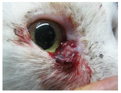
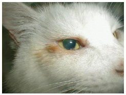
The general systemic condition of the patient was not reflective of suggestive changes of any other pathological condition. Ophthalmology evaluation revealed no abnormalities. Tear production in both the eyes was in the normal range of Schirmer’s I test, (Merck Animal Health, Summit, NJ, USA), fundoscopy results were normal, and the anterior and posterior segments were in perfect condition. The medial region of the right eye’s lower eyelid had an irregular and hemorrhagic mass that was 1 cm in diameter (Figure 1). Differential diagnoses included SCC, hemangiosarcoma, and hemangioma.
Figure 1. A reddish, irregular, and hemorrhagic palpebral mass
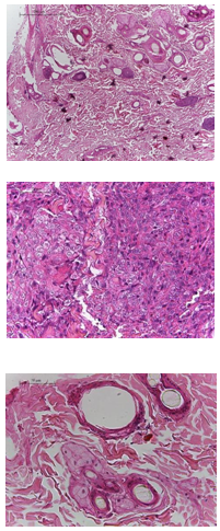
An incisional biopsy of the lesion was obtained under local anesthesia with 2% lidocaine (Virbac, Colombia). The tissue obtained was sent for histopathological analysis in a 10% formaldehyde medium. No treatment was initiated until a histopathological diagnosis was obtained. The histopathology analysis revealed a benign palpebral margin hemangioma (Figures 2–4).
Figures 2–4. Histological slices of the injury. Proliferation of the mesenchymal cells forming a solid mass in the shallow and deep stroma, with discrete delimitation by the connective tissue. A mass with the organization of nests and high neovascularization is seen. The cell contour is not distinguishable, with eosinophilic cytoplasm, oval nuclei, round and elongated, with granular chromatin and two and three nucleoli. Occasional mitosis is seen. No cells are observed in the vessels.
Therapeutic approach
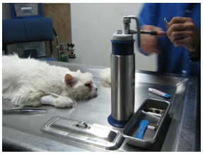
After the pathological result, a cryotherapy session (of approximately 15 min) of the affected region was performed under general anesthesia, with freezing–thawing intervals. A corneal protector (Jager spatula) was used to protect the ocular tissues. A medical liquid nitrogen dermatological applicator was used at −197°C (Figure 5). The decision to perform cryotherapy was based on the risks that conventional blepharoplasty could generate due to the location of the lesion in the corner of the eye, which might have compromised the tear ducts. Postsurgical sequelae showed only slight perilesional inflammation. The only postsurgical medication was anti-inflammatory durinf 4 days.
Figure 5. The patient being prepared for the cryotherapy procedure
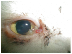
Re-evaluation of the patient a week after the procedure revealed a 90% reduction in the lesion diameter (Figure 6), and overall eye integrity including adequate permeability of the tear ducts as evaluated by the Jones test. A second evaluation was conducted a month post-surgery that revealed total absence of the mass (Figure 7).
Figure 6. Evaluation 1-week post-surgery
Figure 7. Evaluation 1-month post-surgery
Discussion
There are discussions on whether hemangioma can be considered to be a true neoplasm or a vascular malformation. Some points are based on the age of development of the condition. In some species, such as children or foals, these may occur at birth or during early stages of life, which for some authors is suggestive of a vascular malformation (Hargis et al., 1992).
Conclusion
The efficacy in reducing the total diameter of the lesion, rapid results, and limited presentation of adverse effects make cryotherapy an excellent alternative to treat palpebral hemangiomas. It is a minimally invasive and cost-effective tool to treat palpebral neoplasms, and it can replace the conventional techniques such as blepharoplasty that may present with greater risks, such as inadequate edge healing, which can trigger various ocular diseases.
Bibliography
Adzick, NS.; Strome, M.; Gang, D.; Donahoe, PK. Cryotherapy of subglottic hemangioma. Journal of Pedriatric Surgery 1984; 19(4). p. 353-360.
Bickels, J.; Meller, I.; Malawer, MM. The biology and role of cryosurgery in the treatment of bone tumors. En: Malawer MM, Sugarbaker PH. Musculoskeletal cancer surgery treatment of sarcomas and allied diseases. Washington, Kluwer Academic Publisher 2001. p. 135-145.
Dubielzig, RR. Tumors of the eye. En: Meuten DJ. Tumors in domestic animals, 4th ed. Ames, Iowa State, 2002. p. 739-754.
Eltayeb, AA.; Ibrahim, NH.; Moeen, S.; Herdan R. Oral propranolol versus cryotherapy in the management of cutaneous hemangioma in infants and children. Egyptian Journal of Surgery 2016; 35. p. 29-34.
Featherstone, HJ.; Renwick, P.; Heinrich, CL.; Manning, S. Efficacy of lamellar resection, cryotherapy, and adjunctive grafting for the treatment of canine limbal melanoma. Veterinary Ophthalmology 2009; 12, Supplement 1. p. 5-72.
Hargis, AM.; Ihrke, PJ.; Spangler, WL.; Stannard, AA. A retrospective clinicopathologic study of 212 dogs with cutaneous hemangiomas and hemangiosarcomas. Veterinary Pathology 1992; 29. p. 316-328.
Mendirichaga, A.; Vergara, J. Las neoplasias oculares. Argos Portal Veterinaria 2013. p. 1-7.
Michel, S.; Wlotzke, U.; Hohenleutner, U.; Landthaler, M. Laser and cryotherapy of hemangioma in infants in a direct comparison. Hautartz 1998; 49. p. 192-198.
Miller, PE.; Dubielzig, R. Ocular tumors. En: Withrow SA, Vail DM. Small animal clinical oncology, 4th ed. St. Louis, Saunders 2005. p. 686-697.
Newkirk, KM.; Rohrbach, BW. A retrospective study of eyelid tumors from 43 cats. Veterinary Pathology 2009; 46. p. 916-927.
Pirie, CG.; Knollinger, AM.; Thomas, CB.; Dubielzig, RR: Canine conjunctival hemangioma and hemangiosarcoma: a retrospective evaluation of 108 cases (1989-2004). Veterinary Ophthalmology 2006; 9. p. 215-226.
Como citar: Uribe-Castillo, D., Correa-Salgado, R. A. Cryotherapy for Feline Palpebral Hemangioma. Revista Veterinaria y Zootecnia, v. 13, n. 2, p. 73-80, 2019. http://vetzootec.ucaldas.edu.co/index.php/component/content/article?id=278. DOI: 10.17151/vetzo.2019.13.2.7
Esta obra está bajo una Licencia de Creative Commons Reconocimiento CC BY
|


 | Today | 357 |
 | Yesterday | 277 |
 | This week | 1392 |
 | Last week | 3043 |
 | This month | 6496 |
 | Last month | 10358 |
 | All days | 1294131 |
Online Now: 10
Your IP: 18.226.34.215
MOZILLA 5.0,
Today: July 17, 2024
Your IP: 18.226.34.215
MOZILLA 5.0,
Today: July 17, 2024